Volume: 53 Issue: 3 - 2019
| 1. | Cover Pages I - IX |
| ORIGINAL INVESTIGATION | |
| 2. | Evaluation of ischemia-modified albumin levels in acne vulgaris patients Gülhan Gürel, Müjgan Karadöl, Ceylan Bal, Salim Neselioğlu, Emine Çölgeçen doi: 10.4274/turkderm.galenos.2019.71224 Pages 84 - 87 Background and Design: Acne vulgaris (AV) is one of the most common dermatological diseases seen in late childhood and adolescence. The purpose of this study is to evaluate ischemia-modified albumin (IMA) levels in AV, a cutaneous inflammatory disease. Materials and Methods: Seventy-four patients between 15 and 30 years of age with clinically diagnosed AV, and 60 healthy age and sex matched controls without AV were enrolled in the study. Serum IMA levels were measured using the albumin cobalt binding test. The capacity of cobalt to albumin decreases under ischemic conditions. Patient serum is mixed with cobalt chloride and incubated for 5 minutes, during which the cobalt is allowed to bind to the albumin. This is mixed with dithiothreitol (DTT) following incubation, and result in a colored complex with cobalt unbound to albumin for DTT. The resulting colored complex is measured spectrophotometrically at a wavelength of 500 nm. Results: The mean age of the AV group with AV was 18.54 (±3.40), and the mean age of the control group was 17.68 (±3.37). The mean IMA level in the AV patients was 0.82±0.023, compared to 0.80±0.017 in the control group. The difference between the two groups was statistically significant (p<0.001). No significant difference was determined among the AV subgroups in terms of IMA levels. Conclusion: Our search of the Pubmed database revealed no previous studies investigating IMA levels in patients with AV. |
| 3. | Demographic findings of patients diagnosed with pernio and comparison of their vitamin B12, folate and ferritin levels with a control group Ebru Karagün, Sevim Baysak doi: 10.4274/turkderm.galenos.2018.27576 Pages 88 - 92 Background and Design: Pernio is an inflammatory disease that occurs in the acral regions of the body after exposure to cold. The study aimed to investigate the demographic findings of patients diagnosed with pernio and to determine the role of vitamin B12, folate and ferritin values in the pathogenesis of pernio. Materials and Methods: This study was conducted in pernio patients diagnosed at the Ağrı State Hospital, Clinic of Dermatology Outpatient Clinic (Ağrı, Turkey) between September 2015 and April 2016. The healthy control group participants were of similar age and gender. Vitamin B12, folate and ferritin level measurements were compared between the groups. Results: The patient and control groups were similar in age and gender. Vitamin B12 and ferritin levels were significantly lower in the patient group (p<0.05). Folate levels did not differ significantly between the groups (p>0.05). Conclusion: According to the data obtained from our study, when the demographic findings of the pernio patients were compared with those in the literature, the results were found to be similar. In the patients with pernio, it is likely that low Vitamin B12 was involved in the pernio etiology via the mechanism of vasoconstriction/vasodilatation. |
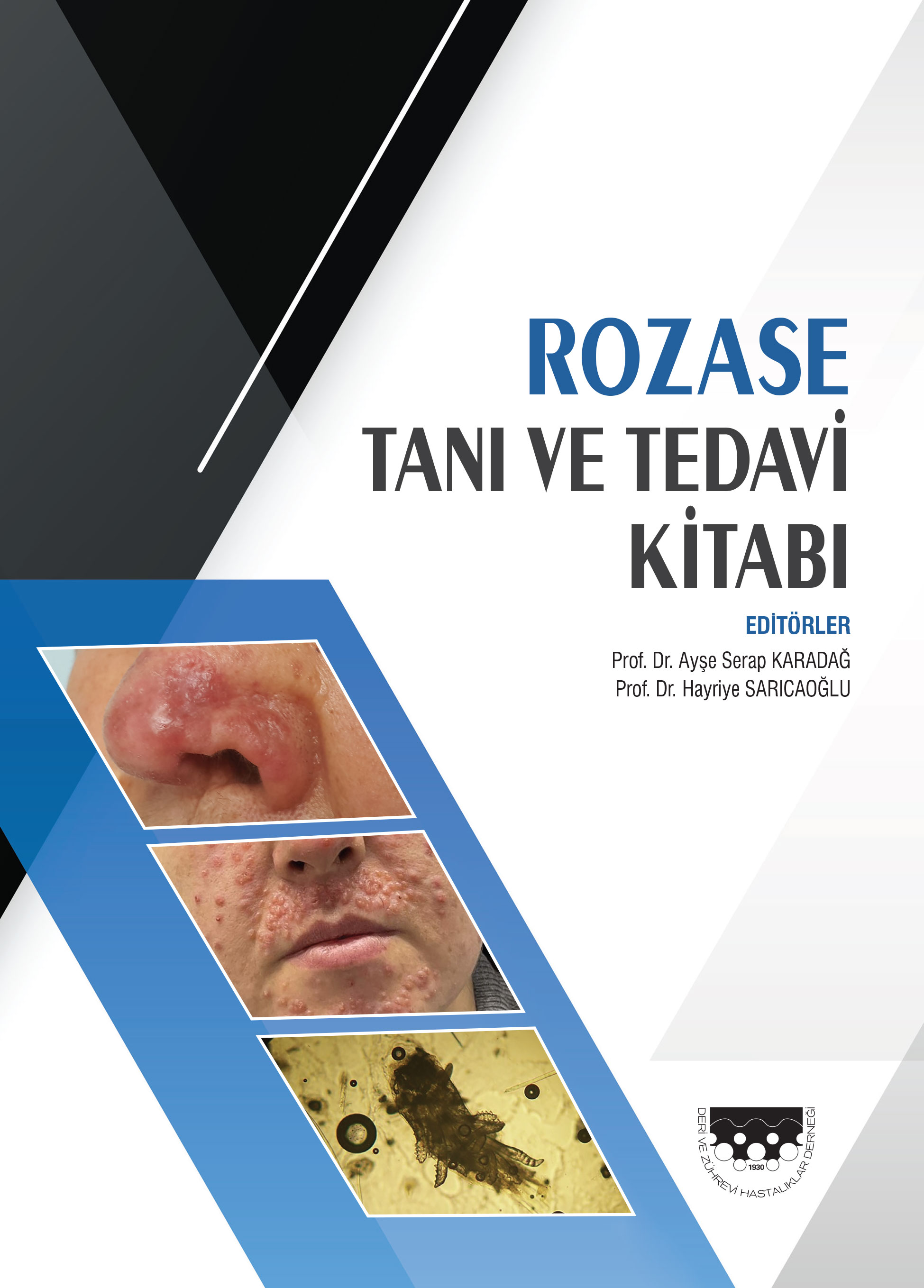
| 4. | Effect of pulsed dye laser therapy on dermatology quality of life in patients with rosacea Serkan Demirkan doi: 10.4274/turkderm.galenos.2019.68726 Pages 93 - 96 Background and Design: Rosacea is a skin disease characterized by erythema, telangiectasia, inflammatory papulopustular eruption and flashing, preferentially affecting the convexities of the face. Among the various treatment options, 595 nm pulsed dye laser (PDL) has been reported to be a good option to treat the erythematotelengiectatic subtype of the disease. Rosacea affects the quality of life negatively, and can cause and clinical findings in patients. The aim of this study was to investigate the effect of PDL on the quality of life in patients with erythematotelengiectatic rosacea. Materials and Methods: Twenty three patients with erythematotelengiectatic rosacea who were treated with three sessions of PDL were asked to complete the Rosacea Investigator Global Assessment scale and Dermatology Life Quality Index (DLQI) questionnaires before and after treatments. Results: The mean pre-treatment DLQI score of the patients was 16.1±0.44, and the mean DLQI score of the patients was 4.3±0.01 after the third treatment. PDL had positive effects on six subcategories of the DLQI. When the patients were asked about the improvement of their disease, there was no significant difference between the first and second PDL applications. However, there was a significant difference between the second and third post-treatment scores. Laser therapy has revolutionized the treatment of erythematotelengiectatic rosacea according to other treatment modalities, and PDL shows a positive effect on the DLQI in patients with erythematotelengiectatic rosacea. Conclusion: PDL is a safe and effective treatment option in erythematotelangiectatic rosacea. It also helps to improve the quality of life in patients. |
| 5. | Erythrasma frequency in patients with interdigital maceration Dursun Türkmen, Gamze Türkoğlu doi: 10.4274/turkderm.galenos.2019.05926 Pages 97 - 100 Background and Design: Erythrasma is a superficial, local, mild and chronic skin infection caused by Corynebacterium minutissimum. Interdigital variant is the most frequent site for bacterial infection of the feet and it is the most common form of erythrasma. It frequently involves moist and closed intertriginous areas together with fissure or squama. The purpose of this study is to research the frequency of erythrasma in patients who refer with interdigital maceration and who are generally considered to have tinea pedis. Materials and Methods: The study was conducted between 15 January 2018 and 15 April 2018. A total of 116 patients who referred with the complaint of interdigital maceration were included in the study. A questionnaire form including demographic information were completed for all patients. All patients were examined with Wood lamp, smears taken were stained with Gram staining and examined under direct microscopy with 20% potassium hydroxide. Results: Of all the 116 patients with interdigital maceration, 97 (83.6%) were diagnosed with erythrasma. Of these 97 erythrasma patients, 66 (68%) were male, 31 (32%) were female and the average age was 48.27±16.29 (minimum age: 20, maximum age: 86 years). Fifteen patients had only Gram staining positive, while 7 patients had only Wood examination positive. Seventy-five patients had both Wood examination and Gram staining positive. Fungal infection was found in direct potassium hydroxide examination of 65 (67%) patients. Conclusion: Erythrasma is frequently seen in patients with maceration. It is important to use Wood light examination and Gram staining together to increase diagnostic accuracy. In addition, it should be kept in mind that erythrasma can be seen with candida and dermatophyte infection when interdigital areas are affected. |
| 6. | The efficacy of adalimumab therapy in refractory hidradenitis suppurativa: Retrospective analysis Meltem Türkmen doi: 10.4274/turkderm.galenos.2019.58295 Pages 101 - 105 Background and Design: Hidradenitis suppurativa (HS) is a painful, chronic, inflammatory skin disease and there is no effective standard method for its treatment. The studies on the successful results of adalimumab in the treatment of HS are increasing and its use is approved by the Ministry of Health in our country. Materials and Methods: The efficacy and side effects of adalimumab were evaluated retrospectively in 9 patients who were diagnosed with resistant HS. Adalimumab was administered 40 mg subcutaneously per week. Patients were followed closely for infections and other side effects. In patients, nodules, abscesses and fistulas were determined at the beginning of the treatment, at the 4th and 12th week of the treatment. The values of global pain scores, life quality index scores and C-reactive protein (CRP) levels recorded at the beginning and at the 12th week of the treatment were evaluated. Results: The ages of 9 patients receiving adalimumab for severe HS were between 33-67 (mean: 50.6), and 7 of the patients were male. The disease duration ranged from 2 to 25 years (mean: 13 years) 5 of the patients according to the Hurley classification was determined as stage 2 and 4 as stage 3. In the 12th week, the number of nodules, abscess and draining fistulas were decreased respectively 60.2%, 69.3% and 52.1%. C-reactive protein levels CRP of 16.3 mg/L at the beginning of treatment were 5.5 mg/L at 12 weeks. Before the treatment, life quality index score was 23.4, and after 12 weeks, the score decreased to 6.8. There was a significant improvement in the quality of life. Pain scores of 6.6±3.4 before the treatment were reduced to 1.5±2.5 in the 12th week of treatment. No side effects were observed during treatment. Coclusion: In this study, adalimumab was evaluated as an effective and well tolerated treatment option in moderate and severe HS. |
| 7. | Nail dermoscopy findings in childhood psoriasis Melike Kibar Öztürk, İlkin Zindancı, Betül Sözeri doi: 10.4274/turkderm.galenos.2019.34392 Pages 106 - 112 Background and Design: Nail dermatoscopy or onychoscopy refers to the examination of the nail unit which consists of the proximal and lateral nail folds, hyponychium, nail plate and nail bed non-invasively. Nail biopsies which require surgical expertise are invasive procedures that are seldom performed by dermatologists in diagnosis of psoriasis. Nail dermatoscopy may offer a distinct advantage by helping avoid nail biopsies in diagnosis of childhood psoriasis. To evaluate the dermatoscopic features of nail psoriasis in a pediatric group of patients and to compare these findings with healty children. Materials and Methods: A total of 45 pediatric patients with nail psoriasis (23 girls, 22 boys) and 45 healthy children were enrolled in the study group. Patients with active psoriatic skin lesions (confirmed by skin biopsy) who had at least one psoriatic nail feature were considered to have nail psoriasis. Psoriasis Area and Severity Index and Nail Psoriasis Severity Index scores are evaluated. Patients with onychomycosis were excluded. Dermatoscopic examination of all nails was performed using a manual dermatoscope with x10 magnification. χ2 test was used. Results: The most frequent dermatoscopic features in the nail bed and matrix were pitting (62.2%), punctate leukonychia (55.5%), haemorrhages (splinter and/or dot) (40%), distal onycholysis (40%), black filamentous structures (35.5%), trachyonychia (33.3%) and salmon patches (26.6%) while a reddish background with sparse dotted vessels (46.6%) and periungual desquamation (48.8%) were the most frequent findings in the proximal and lateral nail folds and in the adjacent skin, respectively. Conclusion: This study highlights the usefulness of nail dermoscopy in helping the diagnosis of nail psoriasis by avoiding an additional nail biopsy in the pediatric population. |
| CASE REPORT | |
| 8. | Spontaneous regression of pediatric extragenital lichen sclerosus: A case report Atiye Oğrum, Pervin Dürer doi: 10.4274/turkderm.galenos.2018.08634 Pages 113 - 115 Lichen sclerosus (LS) is an inflammatory chronic dermatosis with unclear etiology. It can affect all individuals with any age, although the majority of cases occur in prepubertal and perimenopausal periods. The anogenital region is predominantly affected, and the extragenital involvement is uncommon in childhood.Anogenital lesions are classically pruritic and painful white or red patches; however extragenital lesions are generally asymptomatic white papules or patches. The most common sites for extragenital lesions are trunk, neck, and axillae. Most genital LS cases resolve spontaneously during puberty in girls; however we have limited knowledge about the prognosis of extragenital LS in children. Here, we report a case of spontaneous regression of extragenital LS in a 14-year-old girl. |
| 9. | A case of tinea corporis with endothrix type hair involvement Dursun Türkmen doi: 10.4274/turkderm.galenos.2018.10179 Pages 116 - 118 Dermatophytoses are the infections of fungi which can live in dead keratin tissue and affect the stratum corneum of epidermis, nails and hairs. Tinea corporis is a superficial dermatophytosis which is seen in skin parts except hairy skin, beard, hand, foot and inguinal region. This report describes a tinea corporis case with endothrix type of hair involvement. A 26-year-old male patient referred for two indurated plaques on the dorsum of the right hand and the extensor surface of the forearm. A potassium hydroxide (KOH) analysis of lesional skin scrapings was negative, while the KOH analysis of the hairs was positive. The patient responded well to systemic terbinafine therapy. In patients who are considered clinically as tinea, if direct KOH analysis from skin scrapings is negative, examination of the hairs at the lesional area may facilitate diagnosis. |
| 10. | İdiopathic palmoplantar filiform hyperkeratosis succesfully treated with systemic isotretinoin Selma Korkmaz, İjlal Erturan, Başak Filiz, Nermin Karahan, İbrahim Metin Çiriş, Mehmet Yıldırım doi: 10.4274/turkderm.galenos.2018.34466 Pages 119 - 121 Filiform hyperkeratosis is a disease that often involves the palmoplantar region and rarely can involve other parts of the body. It is usually associated with malignancies or systemic diseases, but rarely it can also occur as idiopathic. An 18-year-old woman was admitted to our clinic with the complaints of dermatological skin extensions on her hands and feet for 6 months. She stated that there was no contact of any chemical agent and did not receive any topical or systemic treatment for these complaints. The patient was otherwise healthy and her past medical history and family history were non‐contributory. The systemic examination was evaluated as normal. In dermatologic examination, painful, filiform papules were observed in both palmoplantar regions. Hyperkeratosis and focal regular acanthosis was detected in the skin biopsy from the palmar region of the patient. There was no individual with a similar lesion in in her family, and no pathological findings were found in biochemistry, hemogram and tumor markers. Patient was evaluated as idiopathic palmoplantar filiform like hyperkeratosis with these findings and 20 mg/day isotretinoin treatment was started. It was observed that the lesions almost completely retreat after 2 months of treatment. |
| 11. | Vestibular papillomatosis mimicking genital warts in a pregnant woman Atiye Oğrum, Hatice Yılmaz Doğru doi: 10.4274/turkderm.galenos.2018.47701 Pages 122 - 124 Vestibular papillomatosis (VP) is considered as an anatomical variant of vulva and suggested as the counterpart of pearly penile papules in males. The condition, approximately present in about 1% of women, is characterized by pink, smooth, small papules located around the labia minor and vaginal introitus. Although it usually shows no symptoms; some women suffer from complaints such as itching, burn, sensitivity, and pain. Despite human papillomavirus has been accused in the etiology, no outcomes are found to support this association. The similarity of VP to genital warts leads to unnecessary examinations and treatments, thus causing anxiety in patients. Here, we present a case with 37 weeks of pregnancy, who is planned to undergo cesarean section due to diagnosis of genital warts. |
| DERMATOLOGIST ANSWERS FOR COSMETOLOGY QUESTIONS | |
| 12. | Is Radiofrequency Really Effective in Rejuvenation? Arzu Görgülü Eraslan doi: 10.4274/turkderm.galenos.2019.01058 Pages 125 - 127 Abstract | |


