

Volume: 59 Issue: 1 - 2025
| 1. | Cover Pages I - IV |
| ORIGINAL INVESTIGATION | |
| 2. | Evaluation of medical malpractice claims arising from minimally invasive cosmetic procedures in the context of supreme court decisions Ayţe Kurtuluţ Dereli, Abdülkadir Ýzci, Hülya Cenk, Berna Ţanlý, Kemalettin Acar doi: 10.4274/turkderm.galenos.2025.89238 Pages 1 - 6 Background and Design: This study aims to evaluate the significance of physicians’ obligations under contracts of work in the context of medical malpractice claims related to minimally invasive cosmetic procedures. Additionally, it will address the criteria and methodologies employed in the legal assessment of disputes arising from these procedures, particularly in light of the recent rulings by Supreme Courts. Furthermore, it will emphasize the key considerations from a forensic medical perspective. Materials and Methods: The results of the search conducted using the keywords “botox, aesthetics”, “filling, aesthetics”, “laser, epilation”, “laser, aesthetics”, “laser rejuvenation”, “peeling”, “derma pen”, “mesotherapy”, “platelet-rich plasma”, and “hair transplantation” from the “https: //legalbank.net/arama/mahkeme-kararlari” website revealed that all decisions on medical malpractice as a result of dermatological aesthetic procedures were included in the study. The characteristics of the procedure, the resulting damage, and the trial court and supreme court decisions were evaluated. Results: Seventy-four decisions that met the pre-established screening criteria were identified. Most decisions (83.8%, n=62) were related to laser epilation procedures. The findings revealed that the most frequent locations for the procedures were beauty centers and beauty salons, representing 58.1% of the cases. It was determined that 78.3% (n=58) of the judgments were reversed. Among the reasons for reversal, deficiencies in expert reports were noteworthy. Conclusion: A review of medical malpractice claims has revealed that most dermatological aesthetic procedures and laser epilation applications are associated with beauty centers. The decisions of the supreme courts emphasize the importance of the practitioner's authority, the obligation to inform, the management of complications, and whether the contract of work and the supervision and control responsibility of the responsible manager achieved the promised result. |
| 3. | A comparative analysis of acitretin and methotrexate in the treatment of lichen planus Özge Sevil Karstarlý Bakay, Rahime Ekmekçiođlu doi: 10.4274/turkderm.galenos.2025.94659 Pages 7 - 12 Background and Design: Lichen planus (LP) is a common inflammatory dermatosis affecting people of all ages. Acitretin is one of the firstline systemic treatments; however, certain circumstances limit its use and encourage the search for alternative therapies. We aimed to compare the efficacy and safety of methotrexate as an alternative to acitretin. Materials and Methods: This study retrospectively evaluated the treatment response, clinical characteristics, and demographic features of patients who received methotrexate or acitretin for LP between January 2021-2024. Patients who showed clinical improvement and required continued treatment to maintain control were classified as “clinical responders”. Patients who demonstrated a clinical response and remained clear after treatment discontinuation were classified as in “remission”. Patients whose symptoms did not improve with treatment or who continued to develop new lesions were considered “non-responders”. Results: The study included 66 patients. The mean age of the patients was 53.4±9.6; 47 (71.2%) were female, and 16 (28.8%) were male. Thirty-one (46.9%) patients took methotrexate, and 35 (53.1%) took acitretin. The clinical response rate in patients receiving methotrexate (n=30; 96.7%) was significantly higher than in patients taking acitretin (n=28; 80%) (p<0.05). The predicted treatment response duration did not differ significantly (p>0.05) between the group taking methotrexate (15.9 weeks) and the group taking acitretin (13.8 weeks). There was no statistically significant difference in the number and duration of patients achieving remission and the side effect rate of the treatments (p>0.05). Conclusion: Methotrexate and acitretin are effective and safe options in LP treatment. Multicenter randomized controlled trials are needed to develop treatment guidelines. |
| 4. | Evaluation of difficult alopecia areata cases requiring histopathological confirmation Güldehan Atýţ, Ayţenur Ţam Sarý doi: 10.4274/turkderm.galenos.2025.90768 Pages 13 - 17 Background and Design: Alopecia areata (AA) is a common cause of non-cicatricial hair loss on the scalp or hearing-bearing areas, and diagnosis is typically straightforward. In atypical cases, histopathological examination is suggested. This study aims to determine when dermatologists need histopathological confirmation in AA and which clinical and trichoscopic clues aid diagnosis in atypical cases. Materials and Methods: Patients diagnosed with AA clinically and histopathologically were retrospectively included in the study. Age, gender, duration of disease, localizations of lesions, number of lesions, trichoscopy features, initial diagnosis, extra-scalp involvement, and nail characteristics were recorded. Results: 10 (7.3%) of 137 patients had histopathologic examinations. The mean age of patients was 37.9 (±14.68). Five of the ten patients (50%) were male, and five were female. The mean disease duration was 48.7 (±74.55) months. All the patients suffered from their first episode. Patchy AA (n=9, 39.5%) and cicatricial alopecia (n=7, 30.4%) were the two most frequent initial diagnoses. Loss of follicle ostia (n=7), vellus hairs (n=7), anisotrichosis (n=5), yellow dots (n=5), and erythematous background (n=4) were the five most prevalent features. Only one of the patients (10%) has extra-scalp involvement. Two patients have nail features. Conclusion: We assume that some unexpected trichoscopy features, such as loss of follicle ostium and vellus hair, can be observed in cases with long disease duration, whereas typical trichoscopy features, such as exclamation mark hairs, yellow dots, and black dots, are not always detected in these patients. In some challenging cases, the diagnosis may require histopathological confirmation. |
| CASE REPORT | |
| 5. | Telangiectasia macularis multiplex acquisita: A new entity and literature review Emre Zekey, Mustafa Tosun, Rukiye Yasak Güner, Melih Akyol, Ţeyhmus Kaya doi: 10.4274/turkderm.galenos.2024.20678 Pages 18 - 21 Telangiectasia macularis multiplex acquisita (TMMA) is a very rare entity that has been reported in the Asian population to date. Associations with hepatitis infection and several other systemic diseases have been reported. Its association with hepatitis C infection is a newly designated entity. Here, a 42-year-old Turkish patient with hepatitis C infection who was diagnosed with TMMA is presented, and a literature review is given. Recognition of the characteristic presentation of this rare disease will facilitate the clinician's diagnosis and approach. |
| 6. | Erdheim Chester disease: A case with cutaneous involvement Sera Nur Yücesoy, Güllü Gencebay, Ayţe Mine Önenerk, Tumay Ak, Yusuf Demir, Ýpek Sertbudak, Övgü Aydýn Ülgen, Zekayi Kutlubay doi: 10.4274/turkderm.galenos.2024.34682 Pages 22 - 24 Erdheim-Chester disease (ECD) is a rare multisystem disease characterized by the proliferation of non-Langerhans histiocytes. It is characterized by excessive production and accumulation of histiocytes in multiple tissues and organs. Sites of involvement may include long bones, skin, eyes, lungs, brain, pituitary gland, and additional tissues and organs. The disease course varies depending on the organ and degree of involvement. In this case report, we presented the case of a 54-year-old female patient diagnosed with ECD, manifesting with squamous, enduring plaque lesions on the skin. |
| 7. | Tufted angioma masquerading as a granulomatous condition: A rare case report Aliya Anjum Attar, Navakumar Manickam, Seethalakshmi Ganga vellaisamy, Yamini Mogalingam doi: 10.4274/turkderm.galenos.2024.39129 Pages 25 - 29 Tufted angioma (TA) is an uncommon benign vascular tumor that manifests as erythematous macules, plaques, and nodules and progresses slowly. It is histopathologically characterized by tufts of endothelial cells in a cannonball pattern in the dermis. It primarily affects children and adolescents. A 12-year-old child presented to us with a single, painful, sizeable annular plaque on the right side of his neck that had been there since he was 5 years old. Lupus vulgaris, borderline tuberculoid Hansen, annular elastolytic giant cell granuloma, acquired hemangioma, and benign vascular tumors were all evaluated as differential diagnoses. Ultrasound of the neck revealed an ill-defined hypoechoic region on the right side of the neck with no extension into the deeper plane. TA was diagnosed based on histopathology. We report this case because of its uncommon clinical form of TA, which presented as a single large erythematous annular plaque resembling a granulomatous lesion, which was later confirmed by histological features. |
| LETTER TO THE EDITOR | |
| 8. | Exaggerated subungual fibromas in a patient with tuberous sclerosis complex Dilek Menteţođlu, Ezgi Tosun Yýlmaz, Nilüfer Kök, Selda Pelin Kartal doi: 10.4274/turkderm.galenos.2025.84769 Pages 30 - 31 Abstract | |
| 9. | Acral speckled hypomelanosis: Is there an autoimmune origin? Özge Zorlu, Sevil Karabađ doi: 10.4274/turkderm.galenos.2024.32067 Pages 32 - 34 Abstract | |
| 10. | case of giant cutaneous squamous cell carcinoma mimicking a chronic leg ulcer Handan Merve Erol Mart, Pelin Koçyiđit, Ýncilay Kalay Yýldýzhan, Aylin Okçu Heper, Bengü Nisa Akay doi: 10.4274/turkderm.galenos.2024.73669 Pages 35 - 37 Abstract | |
| BOOK NOTICE | |
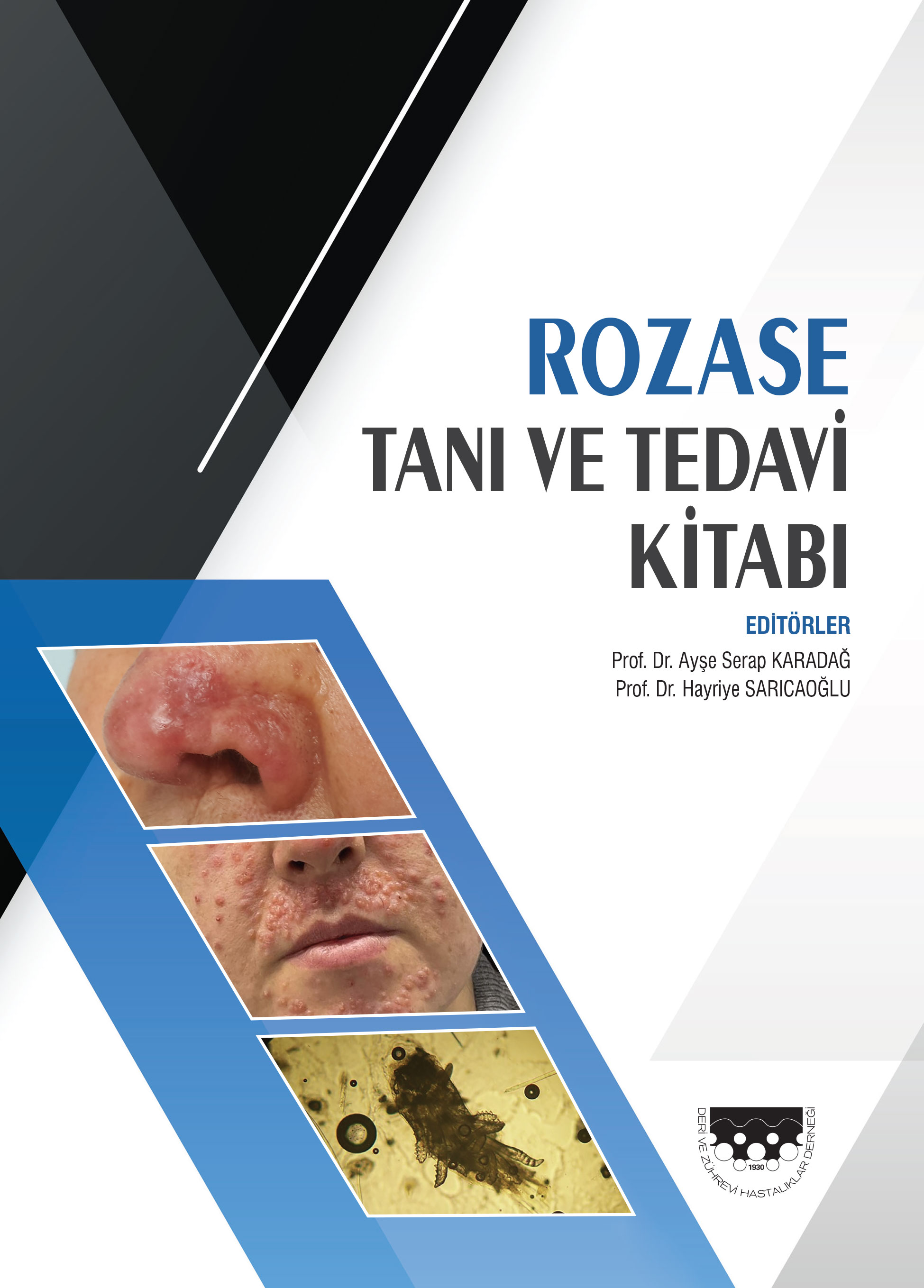
| 11. | Rosacea Diagnosis and Treatment Ayţe Serap Karadađ, Hayriye Sarýcaođlu Page 38 Abstract | |