Volume: 41 Issue: 3 - 2007
| EDITORIAL | |
| 1. | Update on the Treatment of HSV and HPV Infections Ertuğrul H. Aydemir Pages 75 - 76 Abstract | |
| REVIEW ARTICLE | |
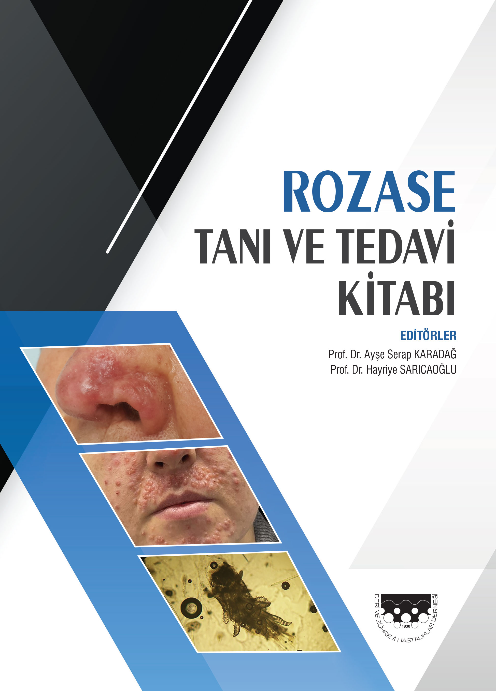
| 2. | The Etiopathogenesis and the new classification system of rosacea Özgür Bakar, Zeynep Demirçay Pages 77 - 80 Rosacea is a chronic, recurrent and an inflammatory disorder of the skin. It is most often characterized by transient or persistent central facial erythema, telangiectases, and often papules and pustules. Although several hypotheses have been documented on the etiopathogenesis of rosacea, the exact cause still remains unknown. Psychogenic, racial, infectious, immunological and vascular theories have been proposed. Recently, a new classification system of rosacea is reported by an expert committe assembled by The National American Rosacea Society. According to this committee, 4 subtypes of rosacea including erythematotelangiectatic, papulopustular, phymatous, and ocular are defined. Current pathogenic theories on rosacea and this new classification system is reviewed in this manuscript. |
| ORIGINAL INVESTIGATION | |
| 3. | Knowledge, attitudes and behaviours of university students related to sun protection Yeşim Kaymak, Ömer Faruk Tekbaş, Işıl Şimşek Pages 81 - 85 Summary: Backgorund and Design: Epidemiologic studies have shown that sun light are the most common environmental factor responsible for the development of cutaneous melanoma, other skin cancers and many other skin diseases. Aim of this study was to investigate the knowledge, attitudes and behaviours of university students related to sun protection. MATERIALS-METHODS: The study was composed of 62 men and 117 women making a total of 179 university students. Subjects were requested to respond to a questionnarie composed of multiple response answers and fill in the blank questiones which was formed by contribution of specialists and literature. Resuts: Most common sources for knowledge about effects of UV light were TV/internet (69.8%), press (52.0%), school (34.1%), family (24.6%) and friends (22.9%), respectively. Subjects responded truly most commonly to vit D synthesis (82.7%), and least to effects on vision (15.6%) as beneficial effects of UV light. Subjects responded truely most commonly to pigmentation (84.9%) and least to allergic reactions (11.7%) as hazardous effects of UV light. Sex and age had no effect on the responses to questions related to knowledge. One hundred nineteen (66.5%) subjects declared tendency to protection against sun, 50 (27.9%) subjects used sun protective agents and 81(45.3%) avoid exposure to sun at dangerous hours (10.00-16.00). CONCLUSION: Results of this study indicate that attitudes and behaviours of university students related to sun protection were not sufficient. |
| 4. | HLA haplotypes in turkish patients with pemphigus and their healthy relatives Mukaddes Kavala, Burçe Can, Sarper Diler, Fatma Oğuz, Emel Önal, Emek Kocatürk Pages 86 - 89 Background and Design: An association between pemphigus vulgaris and certain HLA serotypes that has been found in some ethnic groups suggests the presence of a genetic predisposition for the disease. The aim of this study was to evaluate the antigen frequencies of HLA-A, B, HLA-DR and DQ in patients with pemphigus and their healthy relatives and to adress their association with the disease. MATERIALS-METHODS: HLA class I antigens were determined by standart microcytotoxicity test in 38 patients with pemphigus, 35 healthy relatives, and in control group of 72 individuals while class II antigens were determined by PCR-SSP test in 41 patients with pemphigus, 43 healthy relatives and in 72 controls. RESULTS: HLA class I antigens, A26(10), A28, B44 and HLA class II antigens, DR14 were significantly high in pemphigus patients whereas HLA-A30, A32, B35 and HLA-DR16 were high among controls. In healthy relatives of the patients HLA class I antigens, A26, A28, B57, B62 and HLA class II antigens DR4, DR14, DQ1 were found significantly high while HLA-A29, A30 and HLA-DR3, DR11, DQ5,DQ6 were significantly low. There was no significant association of the other HLA-A,B,DR,DQ antigens with pemphigus. CONCLUSION: Our findings suggest that the occurrence of HLA-A28 and B44 is specific for the Turkish population having an impact in disease susceptibility whereas HLA-A30, A32 and DR16 have a protective role in pemphigus. |
| 5. | The effect of isotretinoin on biotinidase activity Mustafa Kulaç, Ahmet Kahraman, Şemsettin Karaca, Mustafa Serteser Pages 90 - 92 Background and Design: Isotretinoin has many adverse reactions such as cheilitis, xerosis, alopecia, hyperlipidemia, hepatotoxicitity and teratogenicity. Biotinidase is mainly produced in the liver and partial biotinidase deficiency causes dermatological manifestations, seborrheic dermatitis, alopecia etc. Some studies suggests that isotretinoin reduces the serum botinidase activity and some adverse effects might be related this deficiency. In addition, some literatures had reported that the teratogenic findings related hypervitaminosis A has similar characteristics the teratogenic findings related biotinidase deficiency. In this study, we aimed to investigate the effect of isotretinion on biotinidase activity in Turkish patients with acne vulgaris. MATERIAL-METHOD: Thirty patients with acne vulgaris were enrolled to study. The patients had liver function tests, lipid estimations, and biotinidase activity evaluations at the beginning and on the 60th day of treatment with isotretinoin. The same laboratory tests were evaluated in 30 controls only once. RESULTS: Serum biotinidase activity was found to be significantly decreased as compared to the initial values (Initial value: mean: 15.11±9.71 U/l; 60th day: 7.56±5.48 U/l; p<0.0001). A statistically significant elevation of liver enzymes and lipids, except high density lipoprotein, was observed at the end of this study. CONCLUSION: It is suggested that isotretinoin isomers-metabolites act in the liver, resulting in low biotinidase activity. Some dermatological adverse reactions associated isotretinoin therapy such as skin fragility, alopecia might be related partial biotinidase deficieny. |
| 6. | Epidemiologic findings of patients with cutaneous leishmaniasis seen in Antalya Ayşe Akman, Çiçek Durusoy, Deniz Seçkin, Erkan Alpsoy Pages 93 - 96 Background and Design: Cutaneous leishmaniasis is still considered an important health issue in many parts of the world including our country. The disease is endemic in the Southeastern Anatolia region and Çukurova district in the Mediterranean region in Turkey. Herein, we aimed to review the epidemiological data of patients with cutaneous leishmaniasis in Antalya. MATERIALS-METHODS: In this study, patients with cutaneous leishmaniasis, admitted to the Akdeniz University, School of Medicine, Department of Dermatology&Venerology and the Başkent University, School of Medicine, Department of Dermatology, Alanya Hospital between January 2004 and January 2006 were evaluated for their demographical and epidemiological data. RESULTS: Of 20 patients, 10 were women and 10 were men. The age range was between 2 and 40 (mean; 15.05±10.68 years). In the first half of year, the development of the disease was seen in each month. Hovewer, the disease was observed only in August and October in the second half of year. When the case distribution were evaluated in terms of districts where the patiens live, the data are as follows: 8 (40%) in Alanya, 8 (40%) in Gazipaşa, one (5%) in Antalya Center, one (5%) in Serik, one (5%) in Finike, and one (5%) in Kumluca. CONCLUSION: This study showed that patients with cutaneous leishmaniasis were also observed in Antalya Centre. Being near to the endemic regions and having a suitable wheather conditions and humidity, Antalya is likely to be a candidate for an endemic region for cutaneous leishmaniasis. |
| CASE REPORT | |
| 7. | A childhood case with pemphigus herpetiformis Murat Durdu, Soner Uzun, Mehmet Karakaş, Ali Murat Ceyhan, İlhan Tuncer Pages 97 - 100 Pemphigus herpetiformis is a rare variant of pemphigus characterized by a unique clinical phenotype of erythematous or urticarial plaques and vesicles that present in a herpetiform arrangement. Here, we described a 13-year-old-boy who presented with mild pruritic annular erythema and vesicles in a herpetiform arrangement on the trunk and extremities. Mucous membranes were intact. Tzanck smear showed acantholytic cells. Histopathological examination of lesions revealed subcorneal acantholysis and neutrophilic infiltration. Direct immunofluorescence examination demonstrated IgG and C3 deposition on keratinocyte cell surfaces, and indirect immunofluorescence examination showed circulating IgG autoantibodies against keratinocyte cell surfaces at a titer of 1: 20. Based on clinical and laboratory findings, pemphigus herpetiformis was diagnosed. This case is found worth to be presented for both displaying a rare form of pemphigus and for the relatively low incidence of the disease in childhood. |
| 8. | Annular elastolytic giant cell granuloma: Case report Aysun Aktürk, Nilgün Bilen, Metin Yavuz, Kürşat D. Yıldız, Rebiay Kıran Pages 101 - 104 Annuler elastolytic giant cell granuloma (AEGCG) is a rare dermatosis which is characterized by phagocytosis of elastic fibres in dermis histopathologically. It is seen mostly in women as annular plaques located in skin areas exposed to sunlight. Fifty-five year-old female patient, presented to our department with annular plaques that developed on her face. She was diagnosed as AEGCG based on her clinical appearance, histopathological and laboratory findings after exclusion of lupus vulgaris and sarcoidosis. A moderate response in lesions was observed after 9 month with chloroquine phosphate treatment. We have presented our case in order to emphasize the necessity for remembering AEGCG in the differantial diagnoses of granulomatous dermatoses representing itself with annular lesions. |
| 9. | A case of diffuse neonatal hemangiomatosis improving without treatment Şirin Pekcan Yaşar, Zehra Aşiran Serdar, Nurhan Kocayan, Ayşe Tülin Mansur, Ayşe Deniz Akkaya Pages 105 - 107 Diffuse neonatal hemangiomatosis is a rare disorder with poor prognosis that is characterized by cutaneous and visceral hemangiomas, present at birth or develop during neonatal period. Since the mortality rate is as high as 60-95%, treatment with systemic steroids, interferon alpha 2a or partial hepatic resection is often considered, however spontaneous involution is rarely reported. Even if visceral involvement occurs, this may be limited or asymptomatic and so may not always indicate a poor prognosis. Herein, we describe a neonate case with numerous cutaneous hemangiomas and a solitary hepatic hemangioma, complicated with anemia, who showed spontaneous involution of hemangiomas at 8 months. |
| WHAT IS YOUR DIAGNOSIS? | |
| 10. | An erythematous plaque on the chest Şehriyar Nazari Page 108 Abstract | |
| NEW PUBLICATIONS | |
| 11. | Yeni Kitaplar Page 109 Abstract | |
| NEWS | |
| 12. | Kongre Takvimi Page 110 Abstract | |


